濟南維修熱線
13153199508
長沙維修熱線:13873135765
歡迎來到匠仁醫療設備有限公司網站!
濟南維修熱線
13153199508
長沙維修熱線:13873135765

 硬管內窺鏡的維修及介紹
Maintenance and Introduction of Hard Tube Endoscope
關于硬管鏡
Regarding the rigid tube mirror
硬管內窺鏡,主要用于人體表淺及淺層部位自然腔道和通過穿刺開口腔道的病灶診斷和(或)治療,如膀胱鏡、宮腔鏡,在操作中不可彎曲。
Hard tube endoscopes are mainly used for the diagnosis and/or treatment of lesions in superficial and superficial natural cavities of the human body, as well as through puncture opening cavities, such as cystoscopy and hysteroscopy. They cannot be bent during operation.
發展歷史
Development history
內窺鏡是人類窺視自身體內器官的重要工具。古希臘名醫,有著醫藥之父之稱的希波克拉底(Hippocrates,約公元前460-前370)曾描述過一種直腸診視器,該診視器與我們今天所用的器械十分相似。類似的診視器還發現于龐培遺跡(Pompeii,意大利古都,公元79年火山爆發,全城淹沒),這些診視器曾被用于窺視陰道與子宮頸,檢查直腸,并用于檢視耳、鼻內。當時進行這些檢查時利用的是自然光線。內窺鏡的真正發展還是起于近代,一般可將其發展階段分為:硬管式窺鏡、半可屈式內窺鏡、纖維內窺鏡、超聲與電子內窺鏡等階段。硬管式內窺鏡的發展經歷了兩個階段:即開放式硬管內窺鏡階段與含有光學系統的硬管內窺鏡階段。
Endoscope is an important tool for humans to observe their internal organs. Hippocrates, the renowned physician of ancient Greece and known as the father of medicine, described a rectal sight that was very similar to the instruments we use today. Similar examination devices have also been found in the Pompeii ruins (the ancient capital of Italy, where a volcano erupted in 79 AD and the entire city was submerged). These examination devices were once used to observe the vagina and cervix, examine the rectum, and examine the ears and nose. At that time, natural light was used for these inspections. The true development of endoscopes dates back to modern times and can generally be divided into stages such as rigid tube endoscopes, semi flexible endoscopes, fiber endoscopes, ultrasound and electronic endoscopes. The development of rigid tube endoscopes has gone through two stages: the open rigid tube endoscope stage and the rigid tube endoscope stage containing an optical system.
基本結構與工作原理
Basic structure and working principle
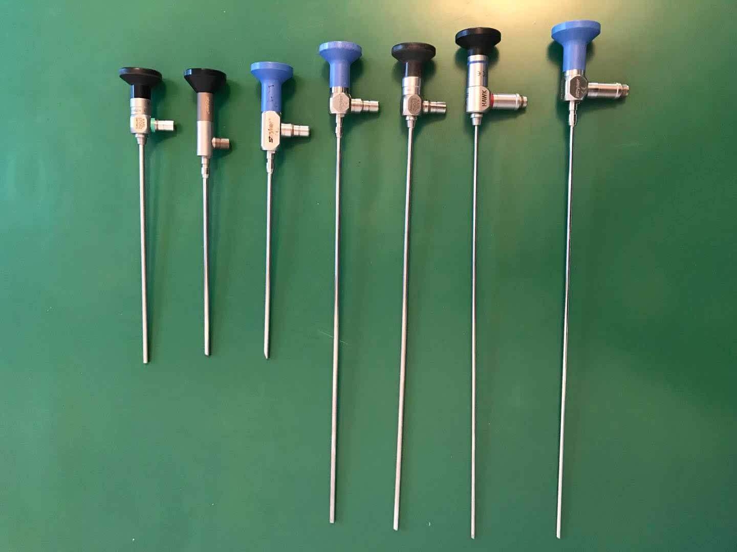
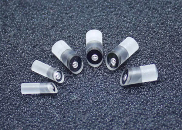
硬管內窺鏡主要由光學成像系統和照明系統組成:光學部分外觀看是一個細長的金屬管子,而里面裝著一個由許多透鏡組成的完整的光學系統。光學成像系統由物鏡系統、轉像系統、目鏡系統三大系統組成。被觀察物經物鏡所成的倒像,通過轉像系統將倒像轉為正像,并傳輸到目鏡,再由目鏡放大后,為人眼所觀察。如圖1所示。為構成不同的視向角,需加入不同的棱鏡。不同用途的內窺鏡根據使用要求制作成不同的外形、外徑、長度,以達到使用所需的要求。照明傳輸系統由光導纖維組成,將冷光源的光經過光導纖維傳輸到內窺鏡前端,照亮被觀察物。
A rigid tube endoscope is mainly composed of an optical imaging system and an illumination system: the optical part is a slender metal tube for external viewing, while inside it is a complete optical system composed of many lenses. The optical imaging system consists of three major systems: the objective lens system, the conversion system, and the eyepiece system. The inverted image of the observed object formed by the objective lens is converted into a positive image through a conversion system and transmitted to the eyepiece. After being magnified by the eyepiece, it is observed by the human eye. As shown in Figure 1. To form different viewing angles, different prisms need to be added. Endoscopes for different purposes are made into different shapes, outer diameters, and lengths according to usage requirements to meet the required requirements. The lighting transmission system is composed of optical fibers, which transmit the light from the cold light source to the front end of the endoscope through the optical fibers, illuminating the observed object.
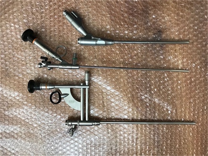
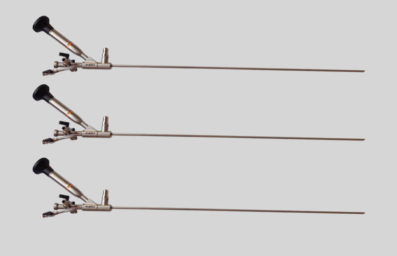
硬式內鏡分類及應用
Classification and application of rigid endoscopes
01
01
膀胱電切鏡
Bladder electrocautery
膀胱電切鏡多用于對膀胱內部的疾病組織進行無創手術,具有風險低、成功率高、手術簡單、愈合快、治愈率高等優點。膀胱電切鏡適用于間質性膀胱炎、濾泡性膀胱炎、腺性膀胱炎、氣性膀胱炎、壞疽性膀胱炎等各種膀胱炎癥的治療。
Bladder electrocautery is commonly used for non-invasive surgery of diseased tissues inside the bladder, with advantages such as low risk, high success rate, simple surgery, fast healing, and high cure rate. Cystoscopy is suitable for the treatment of various bladder inflammations such as interstitial cystitis, follicular cystitis, glandular cystitis, gas cystitis, gangrenous cystitis, etc.
02
02
膀胱鏡
Cystoscopy
膀胱鏡是內窺鏡的一種,外形與尿道探子相似,電鏡鞘、檢查窺鏡、處置和輸尿管插管窺鏡以及鏡芯四部分構成一套,并附有電灼器、剪卡器和活組織檢查鉗等附件。
Cystoscopy is a type of endoscope that resembles a urethral probe in appearance. It consists of four parts: an electron microscope sheath, an inspection scope, a treatment and ureteral catheterization scope, and a core. It is equipped with accessories such as electrocautery, clip clippers, and biopsy forceps.
03
03
腹腔鏡
laparoscope
與電子胃鏡類似,是一種帶有微型攝像頭的器械。腹腔鏡手術與傳統手術相比,具有切口小、痛苦小、恢復快等優點,深受患者的歡迎,尤其是術后瘢痕小、又符合美學要求,青年病人更樂意接受,微創手術是外科發展的總趨勢和追求目標。目前,腹腔鏡手術的金標準是膽囊切除術,一般地說,大部分普通外科的手術,腹腔鏡手術都能完成。如闌尾切除術,胃、十二指腸潰瘍穿孔修補術、疝氣修補術、結腸切除術、脾切除術、腎上腺切除術,還有卵巢囊腫摘除、宮外孕、子宮切除等,隨著腹腔鏡技術的日益完善和腹腔鏡醫生操作水平的提高,幾乎所有的外科手術都能采用這種手術。
Similar to electronic gastroscopy, it is a device with a miniature camera. Compared with traditional surgery, laparoscopic surgery has the advantages of smaller incisions, less pain, and faster recovery, which is highly welcomed by patients. Especially with small postoperative scars and aesthetic requirements, young patients are more willing to accept minimally invasive surgery, which is the overall trend and pursuit goal of surgical development. At present, the gold standard for laparoscopic surgery is cholecystectomy. Generally speaking, most general surgical procedures can be performed using laparoscopic surgery. Such as appendectomy, repair of gastric and duodenal ulcer perforation, hernia repair, colectomy, splenectomy, adrenalectomy, as well as removal of ovarian cysts, ectopic pregnancy, hysterectomy, etc., with the increasing improvement of laparoscopic technology and the improvement of laparoscopic doctors' operating level, almost all surgical procedures can adopt this type of surgery.
04
04
宮腔鏡
Hysteroscopy
宮腔鏡是一項新的、微創性婦科診療技術,可用于診斷、治療和隨訪子宮腔內病變,其實質上是一種纖維光源內窺鏡,包括宮腔鏡、能源系統、光源系統、灌流系統和成像系統。
Hysteroscopy is a new, minimally invasive gynecological diagnostic and therapeutic technique that can be used for the diagnosis, treatment, and follow-up of intrauterine lesions. Essentially, it is a fiber optic endoscope that includes hysteroscopy, energy system, light source system, perfusion system, and imaging system.
05
05
關節鏡
arthroscope
關節鏡是一種觀察關節內部結構的直徑5mm左右的棒狀光學器械,是用于診治關節疾患的內窺鏡。除了對多種運動損傷具有治療作用之外,關節鏡技術在關節炎外科治療中也可大顯身手,發揮重要的作用。關節鏡技術可運用于骨性關節炎、炎癥性關節、色素沉著絨毛結節性滑膜炎、晶體性關節病,感染性關節炎和創傷性關節炎等多種關節炎的診斷和治療。
Arthroscopy is a rod-shaped optical instrument with a diameter of about 5mm used to observe the internal structure of joints, and is an endoscope used for the diagnosis and treatment of joint diseases. In addition to its therapeutic effect on various sports injuries, arthroscopic technology can also play an important role in the surgical treatment of arthritis. Arthroscopy technology can be used for the diagnosis and treatment of various types of arthritis, including osteoarthritis, inflammatory joints, pigmented villonodular synovitis, crystalline arthritis, infectious arthritis, and traumatic arthritis.
06
06
經皮腎鏡
Percutaneous nephrolithotomy
經皮腎鏡技術(PCNL)是腔內泌尿外科手術的一個重要部分。
Percutaneous nephrolithotomy (PCNL) is an important part of endourological surgery.
07
07
前列腺汽化電切鏡
Prostate vaporization electric resection mirror
前列腺汽化電切治療前列腺增生癥被全世界定位前列腺增生治療的金標準。另外,汽化電切鏡還用與尿道腫瘤、膀胱腫瘤的電切以及前列腺癌的姑息性切除治療等。
The treatment of benign prostatic hyperplasia with vaporization and resection of the prostate has been recognized as the gold standard for the treatment of benign prostatic hyperplasia worldwide. In addition, vaporization electrocautery is also used for electrocautery of urethral tumors, bladder tumors, and palliative resection of prostate cancer.
08
08
輸尿管鏡
Ureteroscope
近十幾年來是我國泌尿外科迅猛發展的時期,輸尿管鏡手術是泌尿外科領域發展最快,它是高科技在泌尿外科領域的應用,屬微創診斷、治療的手段,深受廣大病人的歡迎,它具有創傷小、安全,使不少過去要用開放手術的病人免除手術的創傷和痛苦。
In the past decade, China's urology department has experienced rapid development, with ureteroscopy being the fastest growing field. It is a high-tech application in urology, a minimally invasive diagnostic and treatment method, and is highly welcomed by patients. It has the advantages of minimal trauma and safety, allowing many patients who used to undergo open surgery to avoid the trauma and pain of surgery.
維護保養與注意事項
Maintenance and Precautions
隨著使用硬管內窺鏡的范圍擴大,各科醫生使用硬管內窺鏡頻率越來越高。硬管內窺鏡是比較嬌貴的醫療器械,但是只要能夠正確的使用和維護,就可以避免損壞。
With the expansion of the scope of using rigid endoscopes, doctors from various departments are using them more frequently. Hard tube endoscope is a relatively delicate medical device, but as long as it can be used and maintained correctly, damage can be avoided.
維護保養
Maintenance and upkeep
1
one
硬管內窺鏡使用完后應沖洗干凈,把水漬涼干,鏡頭前端有污漬時應用鏡頭紙、棉簽或細紗布輕輕擦拭。
After using the hard tube endoscope, it should be rinsed clean and the water stains should be dried. If there are stains on the front end of the lens, lens paper, cotton swabs, or fine gauze should be gently wiped.
2
two
硬管內窺鏡應有專人專柜保管,放在專用的包裝箱內,內襯柔軟的海綿或聚氨酯泡沫。
The rigid tube endoscope shall be kept in a special cabinet by a specially assigned person, placed in a special packing box, and lined with soft sponge or polyurethane foam.
3
three
所有硬管內窺鏡和手術器械都要碼放整齊,不得交叉重疊放置,確保箱蓋蓋好后,內部的窺鏡和器械不會在搬運時相互撞擊。
All rigid endoscopes and surgical instruments must be neatly stacked, without overlapping or crossing, to ensure that after the box cover is closed, the internal endoscopes and instruments will not collide with each other during transportation.
4
four
取出或放入硬管內窺鏡時,應雙手平托,輕拿輕放。由于內窺鏡的鏡管很薄,受到擠壓、磕碰、折彎、落地等情況就會彎曲變形,導致鏡片破損或光軸偏移而造成圖像不清楚或不能使用,所以從包裝箱中取出或放入硬管內窺鏡時,應雙手平托輕輕地取出或放入,切忌提起一段拽出。
When removing or placing a rigid tube endoscope, it should be held flat with both hands and handled gently. Due to the thin tube of the endoscope, it may bend and deform when subjected to compression, collision, bending, landing, etc., resulting in lens damage or optical axis deviation, causing unclear or unusable images. Therefore, when taking out or placing a hard tube endoscope from the packaging box, it should be gently taken out or placed with both hands flat, and avoid lifting and pulling it out.
5
five
硬管內窺鏡放在托盤等硬質容器內移動時,注意與其他器械分開放置,不要過分顛簸,以免碰撞到窺鏡。
When moving the rigid endoscope in a tray or other hard container, be careful to separate it from other instruments and avoid excessive shaking to avoid colliding with the endoscope.
6
six
包裝箱內應備有干燥劑保持箱內干燥。
There should be a desiccant inside the packaging box to keep it dry.
7
seven
有些硬管內窺鏡不耐高溫高壓,這主要是因為其鏡端封裝用的環氧樹脂膠在高溫高壓下會開膠而造成鏡管內進水,光學系統進水后就會產生視野模糊,所以這類內窺鏡不能用煮沸和高壓蒸汽等高溫高壓的方法消毒。
Some hard tube endoscopes are not resistant to high temperature and high pressure, mainly because the epoxy resin adhesive used for the sealing of the endoscope end will open under high temperature and high pressure, causing water to enter the endoscope tube. After the optical system enters the water, the field of view will be blurred. Therefore, these endoscopes cannot be disinfected by high-temperature and high-pressure methods such as boiling and high-pressure steam.
注意事項
matters needing attention
1
one
在使用其他器械時,尤其是咬合力較大的鉗、剪類器械應注意鏡管的前端不要伸進器械的咬合區內,以免誤傷鏡管。
When using other instruments, especially pliers and scissors with high biting force, attention should be paid to not inserting the front end of the mirror tube into the bite area of the instrument to avoid accidental injury to the mirror tube.
2
two
有些手術窺鏡是在鞘管內使用,在更換其他角度窺鏡或插拔器械時,應注意動作要輕,不可用力過猛。尤其是插拔窺鏡過程中,當遇到阻力拔不動時應仔細查找原因,必要時應連同鞘管一起拔取,不要用蠻力。
Some surgical endoscopes are used inside the sheath. When replacing other angle endoscopes or inserting or removing instruments, attention should be paid to moving gently and not applying too much force. Especially during the process of inserting and removing the endoscope, if there is resistance and it cannot be pulled out, the reason should be carefully investigated. If necessary, the sheath should be pulled out together, and brute force should not be used.
3
three
當窺鏡配合激光汽化、高頻電切、微波等光電技術進行手術時,應注意窺鏡前端與治療點的距離,保證窺鏡前端不被電擊或燒灼。
When the endoscope is used in conjunction with laser vaporization, high-frequency electric cutting, microwave and other optoelectronic technologies for surgery, attention should be paid to the distance between the front end of the endoscope and the treatment point to ensure that the front end of the endoscope is not electrocuted or burned.
4
four
當配合刨削器來切除病變組織時,如耳鼻喉科、骨科的臨床手術中已經廣泛使用,需要控制刀頭的旋轉部分始終在窺鏡的觀察范圍內,在手術范圍較大時,應先停止刀頭轉動,再移動窺鏡,然后在窺鏡監視下移動刀頭,到合適部位后再開機刨削。
When using a scraper to remove diseased tissue, such as in clinical surgeries in otolaryngology and orthopedics, it is necessary to control the rotation of the blade head to always be within the observation range of the endoscope. When the surgical range is large, the rotation of the blade head should be stopped first, and then the endoscope should be moved. Under the supervision of the endoscope, the blade head should be moved to the appropriate location before turning on the scraper.
5
five
當內窺鏡出現圖像模糊、視線不清等狀況時,要及時與廠家或維修機構聯系,切不可繼續盲目使用而造成手術事故。
When the endoscope shows blurry images or unclear vision, it is necessary to promptly contact the manufacturer or maintenance organization, and not continue to blindly use it, which may cause surgical accidents.
常見故障與維修方案
Common faults and maintenance solutions
常見故障
Common faults
1.圖像情況:模糊進水,黑影,灰塵
1. Image condition: Blurred water, black shadows, dust
2.光束:發黃斷絲,灼燒崩缺,亮度不夠
2. Beam: Yellowing and broken wires, burning and chipping, insufficient brightness
3.鏡管:彎曲,斷裂,凹陷,脫落
3. Mirror tube: bent, broken, dented, detached
4.器械通道:磨損崩缺,變形
4. Instrument channel: wear, breakage, deformation
5.物鏡:脫膠,磨損,脫落
5. Objective lens: Degummed, worn, and detached
6.光錐:灼燒發黑,發黃,脫落缺失
6. Light cone: blackened, yellowed, and missing due to burning
7.鏡橋組件:脫落,卡口斷
7. Mirror bridge component: detached, clip broken
8.目鏡保護片:破裂,崩缺,脫落缺失
8. Eyepiece protective film: rupture, breakage, detachment or missing
9.目鏡罩:破裂,崩缺
9. Eyepiece cover: ruptured, broken
維修方案
Repair plan
1.更換內部光學部件
1. Replace internal optical components
2.更換光束
2. Replace the beam
3.更換鏡管
3. Replace the mirror tube
4.更換器械通道
4. Replace the instrument channel
5.更換物鏡
5. Replace the objective lens
6.更換光錐
6. Replace the light cone
7.更換鏡橋組件
7. Replace the mirror bridge component
8.更換目鏡保護片
8. Replace the eyepiece protective film
9.更換目鏡罩
9. Replace the eyepiece cover
本文由 內窺鏡維修 友情奉獻.更多有關的知識請點擊 http://www.zfmm400.com 真誠的態度.為您提供為全面的服務.更多有關的知識我們將會陸續向大家奉獻.敬請期待.
This article is a friendly contribution from endoscope repair. For more related knowledge, please click http://www.zfmm400.com Sincere attitude. We provide you with comprehensive services. We will gradually contribute more relevant knowledge to everyone. Please stay tuned
硬管內窺鏡的維修及介紹
Maintenance and Introduction of Hard Tube Endoscope
關于硬管鏡
Regarding the rigid tube mirror
硬管內窺鏡,主要用于人體表淺及淺層部位自然腔道和通過穿刺開口腔道的病灶診斷和(或)治療,如膀胱鏡、宮腔鏡,在操作中不可彎曲。
Hard tube endoscopes are mainly used for the diagnosis and/or treatment of lesions in superficial and superficial natural cavities of the human body, as well as through puncture opening cavities, such as cystoscopy and hysteroscopy. They cannot be bent during operation.
發展歷史
Development history
內窺鏡是人類窺視自身體內器官的重要工具。古希臘名醫,有著醫藥之父之稱的希波克拉底(Hippocrates,約公元前460-前370)曾描述過一種直腸診視器,該診視器與我們今天所用的器械十分相似。類似的診視器還發現于龐培遺跡(Pompeii,意大利古都,公元79年火山爆發,全城淹沒),這些診視器曾被用于窺視陰道與子宮頸,檢查直腸,并用于檢視耳、鼻內。當時進行這些檢查時利用的是自然光線。內窺鏡的真正發展還是起于近代,一般可將其發展階段分為:硬管式窺鏡、半可屈式內窺鏡、纖維內窺鏡、超聲與電子內窺鏡等階段。硬管式內窺鏡的發展經歷了兩個階段:即開放式硬管內窺鏡階段與含有光學系統的硬管內窺鏡階段。
Endoscope is an important tool for humans to observe their internal organs. Hippocrates, the renowned physician of ancient Greece and known as the father of medicine, described a rectal sight that was very similar to the instruments we use today. Similar examination devices have also been found in the Pompeii ruins (the ancient capital of Italy, where a volcano erupted in 79 AD and the entire city was submerged). These examination devices were once used to observe the vagina and cervix, examine the rectum, and examine the ears and nose. At that time, natural light was used for these inspections. The true development of endoscopes dates back to modern times and can generally be divided into stages such as rigid tube endoscopes, semi flexible endoscopes, fiber endoscopes, ultrasound and electronic endoscopes. The development of rigid tube endoscopes has gone through two stages: the open rigid tube endoscope stage and the rigid tube endoscope stage containing an optical system.
基本結構與工作原理
Basic structure and working principle
硬管內窺鏡主要由光學成像系統和照明系統組成:光學部分外觀看是一個細長的金屬管子,而里面裝著一個由許多透鏡組成的完整的光學系統。光學成像系統由物鏡系統、轉像系統、目鏡系統三大系統組成。被觀察物經物鏡所成的倒像,通過轉像系統將倒像轉為正像,并傳輸到目鏡,再由目鏡放大后,為人眼所觀察。如圖1所示。為構成不同的視向角,需加入不同的棱鏡。不同用途的內窺鏡根據使用要求制作成不同的外形、外徑、長度,以達到使用所需的要求。照明傳輸系統由光導纖維組成,將冷光源的光經過光導纖維傳輸到內窺鏡前端,照亮被觀察物。
A rigid tube endoscope is mainly composed of an optical imaging system and an illumination system: the optical part is a slender metal tube for external viewing, while inside it is a complete optical system composed of many lenses. The optical imaging system consists of three major systems: the objective lens system, the conversion system, and the eyepiece system. The inverted image of the observed object formed by the objective lens is converted into a positive image through a conversion system and transmitted to the eyepiece. After being magnified by the eyepiece, it is observed by the human eye. As shown in Figure 1. To form different viewing angles, different prisms need to be added. Endoscopes for different purposes are made into different shapes, outer diameters, and lengths according to usage requirements to meet the required requirements. The lighting transmission system is composed of optical fibers, which transmit the light from the cold light source to the front end of the endoscope through the optical fibers, illuminating the observed object.
硬式內鏡分類及應用
Classification and application of rigid endoscopes
01
01
膀胱電切鏡
Bladder electrocautery
膀胱電切鏡多用于對膀胱內部的疾病組織進行無創手術,具有風險低、成功率高、手術簡單、愈合快、治愈率高等優點。膀胱電切鏡適用于間質性膀胱炎、濾泡性膀胱炎、腺性膀胱炎、氣性膀胱炎、壞疽性膀胱炎等各種膀胱炎癥的治療。
Bladder electrocautery is commonly used for non-invasive surgery of diseased tissues inside the bladder, with advantages such as low risk, high success rate, simple surgery, fast healing, and high cure rate. Cystoscopy is suitable for the treatment of various bladder inflammations such as interstitial cystitis, follicular cystitis, glandular cystitis, gas cystitis, gangrenous cystitis, etc.
02
02
膀胱鏡
Cystoscopy
膀胱鏡是內窺鏡的一種,外形與尿道探子相似,電鏡鞘、檢查窺鏡、處置和輸尿管插管窺鏡以及鏡芯四部分構成一套,并附有電灼器、剪卡器和活組織檢查鉗等附件。
Cystoscopy is a type of endoscope that resembles a urethral probe in appearance. It consists of four parts: an electron microscope sheath, an inspection scope, a treatment and ureteral catheterization scope, and a core. It is equipped with accessories such as electrocautery, clip clippers, and biopsy forceps.
03
03
腹腔鏡
laparoscope
與電子胃鏡類似,是一種帶有微型攝像頭的器械。腹腔鏡手術與傳統手術相比,具有切口小、痛苦小、恢復快等優點,深受患者的歡迎,尤其是術后瘢痕小、又符合美學要求,青年病人更樂意接受,微創手術是外科發展的總趨勢和追求目標。目前,腹腔鏡手術的金標準是膽囊切除術,一般地說,大部分普通外科的手術,腹腔鏡手術都能完成。如闌尾切除術,胃、十二指腸潰瘍穿孔修補術、疝氣修補術、結腸切除術、脾切除術、腎上腺切除術,還有卵巢囊腫摘除、宮外孕、子宮切除等,隨著腹腔鏡技術的日益完善和腹腔鏡醫生操作水平的提高,幾乎所有的外科手術都能采用這種手術。
Similar to electronic gastroscopy, it is a device with a miniature camera. Compared with traditional surgery, laparoscopic surgery has the advantages of smaller incisions, less pain, and faster recovery, which is highly welcomed by patients. Especially with small postoperative scars and aesthetic requirements, young patients are more willing to accept minimally invasive surgery, which is the overall trend and pursuit goal of surgical development. At present, the gold standard for laparoscopic surgery is cholecystectomy. Generally speaking, most general surgical procedures can be performed using laparoscopic surgery. Such as appendectomy, repair of gastric and duodenal ulcer perforation, hernia repair, colectomy, splenectomy, adrenalectomy, as well as removal of ovarian cysts, ectopic pregnancy, hysterectomy, etc., with the increasing improvement of laparoscopic technology and the improvement of laparoscopic doctors' operating level, almost all surgical procedures can adopt this type of surgery.
04
04
宮腔鏡
Hysteroscopy
宮腔鏡是一項新的、微創性婦科診療技術,可用于診斷、治療和隨訪子宮腔內病變,其實質上是一種纖維光源內窺鏡,包括宮腔鏡、能源系統、光源系統、灌流系統和成像系統。
Hysteroscopy is a new, minimally invasive gynecological diagnostic and therapeutic technique that can be used for the diagnosis, treatment, and follow-up of intrauterine lesions. Essentially, it is a fiber optic endoscope that includes hysteroscopy, energy system, light source system, perfusion system, and imaging system.
05
05
關節鏡
arthroscope
關節鏡是一種觀察關節內部結構的直徑5mm左右的棒狀光學器械,是用于診治關節疾患的內窺鏡。除了對多種運動損傷具有治療作用之外,關節鏡技術在關節炎外科治療中也可大顯身手,發揮重要的作用。關節鏡技術可運用于骨性關節炎、炎癥性關節、色素沉著絨毛結節性滑膜炎、晶體性關節病,感染性關節炎和創傷性關節炎等多種關節炎的診斷和治療。
Arthroscopy is a rod-shaped optical instrument with a diameter of about 5mm used to observe the internal structure of joints, and is an endoscope used for the diagnosis and treatment of joint diseases. In addition to its therapeutic effect on various sports injuries, arthroscopic technology can also play an important role in the surgical treatment of arthritis. Arthroscopy technology can be used for the diagnosis and treatment of various types of arthritis, including osteoarthritis, inflammatory joints, pigmented villonodular synovitis, crystalline arthritis, infectious arthritis, and traumatic arthritis.
06
06
經皮腎鏡
Percutaneous nephrolithotomy
經皮腎鏡技術(PCNL)是腔內泌尿外科手術的一個重要部分。
Percutaneous nephrolithotomy (PCNL) is an important part of endourological surgery.
07
07
前列腺汽化電切鏡
Prostate vaporization electric resection mirror
前列腺汽化電切治療前列腺增生癥被全世界定位前列腺增生治療的金標準。另外,汽化電切鏡還用與尿道腫瘤、膀胱腫瘤的電切以及前列腺癌的姑息性切除治療等。
The treatment of benign prostatic hyperplasia with vaporization and resection of the prostate has been recognized as the gold standard for the treatment of benign prostatic hyperplasia worldwide. In addition, vaporization electrocautery is also used for electrocautery of urethral tumors, bladder tumors, and palliative resection of prostate cancer.
08
08
輸尿管鏡
Ureteroscope
近十幾年來是我國泌尿外科迅猛發展的時期,輸尿管鏡手術是泌尿外科領域發展最快,它是高科技在泌尿外科領域的應用,屬微創診斷、治療的手段,深受廣大病人的歡迎,它具有創傷小、安全,使不少過去要用開放手術的病人免除手術的創傷和痛苦。
In the past decade, China's urology department has experienced rapid development, with ureteroscopy being the fastest growing field. It is a high-tech application in urology, a minimally invasive diagnostic and treatment method, and is highly welcomed by patients. It has the advantages of minimal trauma and safety, allowing many patients who used to undergo open surgery to avoid the trauma and pain of surgery.
維護保養與注意事項
Maintenance and Precautions
隨著使用硬管內窺鏡的范圍擴大,各科醫生使用硬管內窺鏡頻率越來越高。硬管內窺鏡是比較嬌貴的醫療器械,但是只要能夠正確的使用和維護,就可以避免損壞。
With the expansion of the scope of using rigid endoscopes, doctors from various departments are using them more frequently. Hard tube endoscope is a relatively delicate medical device, but as long as it can be used and maintained correctly, damage can be avoided.
維護保養
Maintenance and upkeep
1
one
硬管內窺鏡使用完后應沖洗干凈,把水漬涼干,鏡頭前端有污漬時應用鏡頭紙、棉簽或細紗布輕輕擦拭。
After using the hard tube endoscope, it should be rinsed clean and the water stains should be dried. If there are stains on the front end of the lens, lens paper, cotton swabs, or fine gauze should be gently wiped.
2
two
硬管內窺鏡應有專人專柜保管,放在專用的包裝箱內,內襯柔軟的海綿或聚氨酯泡沫。
The rigid tube endoscope shall be kept in a special cabinet by a specially assigned person, placed in a special packing box, and lined with soft sponge or polyurethane foam.
3
three
所有硬管內窺鏡和手術器械都要碼放整齊,不得交叉重疊放置,確保箱蓋蓋好后,內部的窺鏡和器械不會在搬運時相互撞擊。
All rigid endoscopes and surgical instruments must be neatly stacked, without overlapping or crossing, to ensure that after the box cover is closed, the internal endoscopes and instruments will not collide with each other during transportation.
4
four
取出或放入硬管內窺鏡時,應雙手平托,輕拿輕放。由于內窺鏡的鏡管很薄,受到擠壓、磕碰、折彎、落地等情況就會彎曲變形,導致鏡片破損或光軸偏移而造成圖像不清楚或不能使用,所以從包裝箱中取出或放入硬管內窺鏡時,應雙手平托輕輕地取出或放入,切忌提起一段拽出。
When removing or placing a rigid tube endoscope, it should be held flat with both hands and handled gently. Due to the thin tube of the endoscope, it may bend and deform when subjected to compression, collision, bending, landing, etc., resulting in lens damage or optical axis deviation, causing unclear or unusable images. Therefore, when taking out or placing a hard tube endoscope from the packaging box, it should be gently taken out or placed with both hands flat, and avoid lifting and pulling it out.
5
five
硬管內窺鏡放在托盤等硬質容器內移動時,注意與其他器械分開放置,不要過分顛簸,以免碰撞到窺鏡。
When moving the rigid endoscope in a tray or other hard container, be careful to separate it from other instruments and avoid excessive shaking to avoid colliding with the endoscope.
6
six
包裝箱內應備有干燥劑保持箱內干燥。
There should be a desiccant inside the packaging box to keep it dry.
7
seven
有些硬管內窺鏡不耐高溫高壓,這主要是因為其鏡端封裝用的環氧樹脂膠在高溫高壓下會開膠而造成鏡管內進水,光學系統進水后就會產生視野模糊,所以這類內窺鏡不能用煮沸和高壓蒸汽等高溫高壓的方法消毒。
Some hard tube endoscopes are not resistant to high temperature and high pressure, mainly because the epoxy resin adhesive used for the sealing of the endoscope end will open under high temperature and high pressure, causing water to enter the endoscope tube. After the optical system enters the water, the field of view will be blurred. Therefore, these endoscopes cannot be disinfected by high-temperature and high-pressure methods such as boiling and high-pressure steam.
注意事項
matters needing attention
1
one
在使用其他器械時,尤其是咬合力較大的鉗、剪類器械應注意鏡管的前端不要伸進器械的咬合區內,以免誤傷鏡管。
When using other instruments, especially pliers and scissors with high biting force, attention should be paid to not inserting the front end of the mirror tube into the bite area of the instrument to avoid accidental injury to the mirror tube.
2
two
有些手術窺鏡是在鞘管內使用,在更換其他角度窺鏡或插拔器械時,應注意動作要輕,不可用力過猛。尤其是插拔窺鏡過程中,當遇到阻力拔不動時應仔細查找原因,必要時應連同鞘管一起拔取,不要用蠻力。
Some surgical endoscopes are used inside the sheath. When replacing other angle endoscopes or inserting or removing instruments, attention should be paid to moving gently and not applying too much force. Especially during the process of inserting and removing the endoscope, if there is resistance and it cannot be pulled out, the reason should be carefully investigated. If necessary, the sheath should be pulled out together, and brute force should not be used.
3
three
當窺鏡配合激光汽化、高頻電切、微波等光電技術進行手術時,應注意窺鏡前端與治療點的距離,保證窺鏡前端不被電擊或燒灼。
When the endoscope is used in conjunction with laser vaporization, high-frequency electric cutting, microwave and other optoelectronic technologies for surgery, attention should be paid to the distance between the front end of the endoscope and the treatment point to ensure that the front end of the endoscope is not electrocuted or burned.
4
four
當配合刨削器來切除病變組織時,如耳鼻喉科、骨科的臨床手術中已經廣泛使用,需要控制刀頭的旋轉部分始終在窺鏡的觀察范圍內,在手術范圍較大時,應先停止刀頭轉動,再移動窺鏡,然后在窺鏡監視下移動刀頭,到合適部位后再開機刨削。
When using a scraper to remove diseased tissue, such as in clinical surgeries in otolaryngology and orthopedics, it is necessary to control the rotation of the blade head to always be within the observation range of the endoscope. When the surgical range is large, the rotation of the blade head should be stopped first, and then the endoscope should be moved. Under the supervision of the endoscope, the blade head should be moved to the appropriate location before turning on the scraper.
5
five
當內窺鏡出現圖像模糊、視線不清等狀況時,要及時與廠家或維修機構聯系,切不可繼續盲目使用而造成手術事故。
When the endoscope shows blurry images or unclear vision, it is necessary to promptly contact the manufacturer or maintenance organization, and not continue to blindly use it, which may cause surgical accidents.
常見故障與維修方案
Common faults and maintenance solutions
常見故障
Common faults
1.圖像情況:模糊進水,黑影,灰塵
1. Image condition: Blurred water, black shadows, dust
2.光束:發黃斷絲,灼燒崩缺,亮度不夠
2. Beam: Yellowing and broken wires, burning and chipping, insufficient brightness
3.鏡管:彎曲,斷裂,凹陷,脫落
3. Mirror tube: bent, broken, dented, detached
4.器械通道:磨損崩缺,變形
4. Instrument channel: wear, breakage, deformation
5.物鏡:脫膠,磨損,脫落
5. Objective lens: Degummed, worn, and detached
6.光錐:灼燒發黑,發黃,脫落缺失
6. Light cone: blackened, yellowed, and missing due to burning
7.鏡橋組件:脫落,卡口斷
7. Mirror bridge component: detached, clip broken
8.目鏡保護片:破裂,崩缺,脫落缺失
8. Eyepiece protective film: rupture, breakage, detachment or missing
9.目鏡罩:破裂,崩缺
9. Eyepiece cover: ruptured, broken
維修方案
Repair plan
1.更換內部光學部件
1. Replace internal optical components
2.更換光束
2. Replace the beam
3.更換鏡管
3. Replace the mirror tube
4.更換器械通道
4. Replace the instrument channel
5.更換物鏡
5. Replace the objective lens
6.更換光錐
6. Replace the light cone
7.更換鏡橋組件
7. Replace the mirror bridge component
8.更換目鏡保護片
8. Replace the eyepiece protective film
9.更換目鏡罩
9. Replace the eyepiece cover
本文由 內窺鏡維修 友情奉獻.更多有關的知識請點擊 http://www.zfmm400.com 真誠的態度.為您提供為全面的服務.更多有關的知識我們將會陸續向大家奉獻.敬請期待.
This article is a friendly contribution from endoscope repair. For more related knowledge, please click http://www.zfmm400.com Sincere attitude. We provide you with comprehensive services. We will gradually contribute more relevant knowledge to everyone. Please stay tuned
 維修項目
維修項目 聯系我們
聯系我們 公司:匠仁醫療設備有限公司
  新聞資訊
新聞資訊微信公眾號

樊經理:13153199508 李經理:13873135765
公司地址:山東省濟南市槐蔭區美里東路3000號德邁國際信息產業園6號樓101-2室 湖南省長沙市雨花區勞動東路820號恒大綠洲14棟2409室
備案號:魯ICP備2023027194號-1 魯公網安備 37010402441281號